Our ability to “see” starts when light reflects off an object at which we are looking and enters the eye. As it enters the eye, the light is unfocused. The first step in seeing is to focus the light rays onto the retina, which is the light sensitive layer found inside the eye. Once the light is focused, it stimulates cells to send millions of electrochemical impulses along the optic nerve to the brain. The portion of the brain at the back of the head interprets the impulses, enabling us to see the object.
Light, refraction and its importance
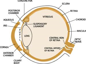
Light entering the eye is first bent, or refracted, by the cornea — the clear window on the outer front surface of the eyeball. The cornea provides most of the eye’s optical power or light-bending ability.
After the light passes through the cornea, it is bent again — to a more finely adjusted focus — by the crystalline lens inside the eye. The lens focuses the light on the retina. This is achieved by the ciliary muscles in the eye changing the shape of the lens, bending or flattening it to focus the light rays on the retina.
This adjustment in the lens, known as accommodation, is necessary for bringing near and far objects into focus. The process of bending light to produce a focused image on the retina is called “refraction”. Ideally, the light is “refracted,” or redirected, in such a manner that the rays are focused into a precise image on the retina.
Most vision problems occur because of an error in how our eyes refract light. In nearsightedness (myopia), the light rays form an image in front of the retina. In farsightedness (hypermetropia), the rays focus behind the retina. In astigmatism, the curvature of the cornea is irregular, causing light rays to focus to more than one place so that a single clear image cannot be formed on the retina, resulting in blurred vision. As we age, we find reading or performing close-up activities more difficult. This condition is called presbyopia, and results from the crystalline lens being less flexible, and therefore less able to bend light.
Since changing the apparent refraction of the eye is relatively easy through the use of corrective spectacle or contact lenses, many of the conditions that contribute to unclear vision can be readily corrected.
How do we make sense of light?
Sensory interpretation
Even with the light focused on the retina, the process of seeing is not complete. For one thing, the image is inverted, or upside down. Light from the various “pieces” of the object being observed stimulate nerve endings — photoreceptors or cells sensitive to light — in the retina.
Rods and cones
Two types of receptors — rods and cones — are present. Rods are mainly found in the peripheral retina and enable us to see in dim light and to detect peripheral motion. They are primarily responsible for night vision and visual orientation. Cones are principally found in the central retina and provide detailed vision for such tasks as reading or distinguishing distant objects. They also are necessary for color detection. These photoreceptors convert light to electrochemical impulses that are transmitted via the nerves to the brain.
Millions of impulses travel along the nerve fibers of the optic nerve at the back of the eye, eventually arriving at the visual cortex of the brain, located at the back of the head. Here, the electrochemical impulses are unscrambled and interpreted. The image is re-inverted so that we see the object the right way up. This “sensory” part of seeing is much more complex than the refractive part — and therefore is much more difficult to influence accurately.